Regulatory Genomics and Systems Biology

News & updates

Intrinsically Disordered Segments Affect Protein Half-Life in the Cell and during Evolution

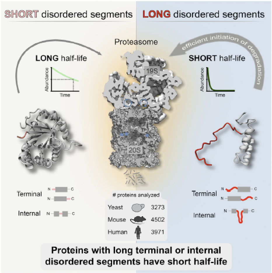
Precise control of protein turnover is essential for cellular homeostasis. The ubiquitin-proteasome system is well established as a major regulator of protein degradation, but an understanding of how inherent structural features influence the lifetimes of proteins is lacking. We report that yeast, mouse, and human proteins with terminal or internal intrinsically disordered segments have significantly shorter half-lives than proteins without these features. The lengths of the disordered segments that affect protein half-life are compatible with the structure of the proteasome. Divergence in terminal and internal disordered segments in yeast proteins originating from gene duplication leads to significantly altered half-life. Many paralogs that are affected by such changes participate in signaling, where altered protein half-life will directly impact cellular processes and function. Thus, natural variation in the length and position of disordered segments may affect protein half-life and could serve as an underappreciated source of genetic variation with important phenotypic consequences. The paper by Robin van der Lee et al can be found here.
Asymmetric mRNA localization contributes to fidelity and sensitivity of spatially localized systems

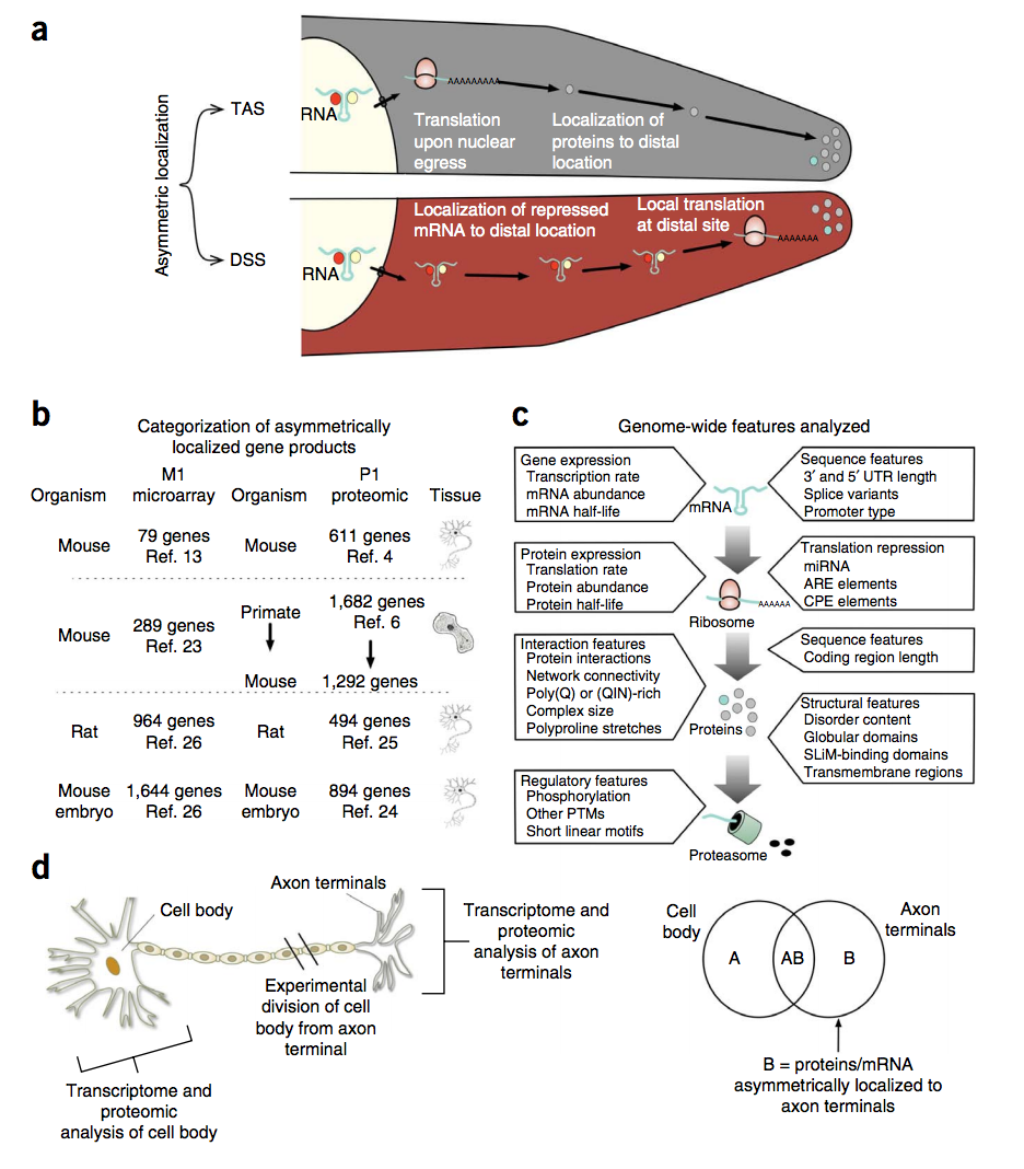
Although many proteins are localized after translation, asymmetric protein distribution is also achieved by translation after mRNA localization. Why are certain mRNA transported to a distal location and translated on-site? Here we undertake a systematic, genome-scale study of asymmetrically distributed protein and mRNA in mammalian cells. Our findings suggest that asymmetric protein distribution by mRNA localization enhances interaction fidelity and signaling sensitivity. Proteins synthesized at distal locations frequently contain intrinsically disordered segments. These regions are generally rich in assembly-promoting modules and are often regulated by post-translational modifications. Such proteins are tightly regulated but display distinct temporal dynamics upon stimulation with growth factors. Thus, proteins synthesized on-site may rapidly alter proteome composition and act as dynamically regulated scaffolds to promote the formation of reversible cellular assemblies. Our observations are consistent across multiple mammalian species, cell types and developmental stages, suggesting that localized translation is a recurring feature of cell signaling and regulation. The paper by Robert Weatheritt et al can be found here.

A Million Peptide Motifs for the Molecular Biologist

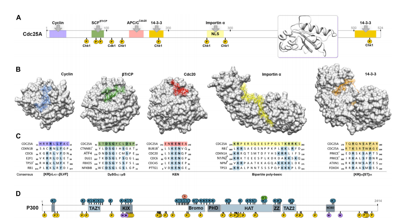
A molecular description of functional modules in the cell is the focus of many high-throughput studies in the postgenomic era. A large portion of biomolecular interactions in virtually all cellular processes is mediated by compact interaction modules, referred to as peptide motifs. Such motifs are typically less than ten residues in length, occur within intrinsically disordered regions, and are recognized and/or posttranslationally modified by structured domains of the interacting partner. In this review, we suggest that there might be over a million instances of peptide motifs in the human proteome. While this staggering number suggests that peptide motifs are numerous and the most understudied functional module in the cell, it also holds great opportunities for new discoveries. The review can be found here.

Use of Host-like Peptide Motifs in Viral Proteins Is a Prevalent Strategy in Host-Virus Interactions

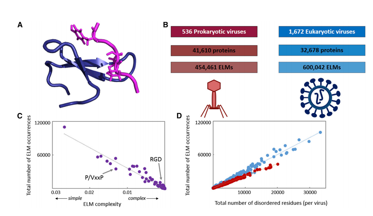
Viruses interact extensively with host proteins, but the mechanisms controlling these interactions are not well understood. We present a comprehensive analysis of eukaryotic linear motifs (ELMs) in 2,208 viral genomes and reveal that viruses exploit molecular mimicry of host-like ELMs to possibly assist in host-virus interactions. Using a statistical genomics approach, we identify a large number of potentially functional ELMs and observe that the occurrence of ELMs is often evolutionarily conserved but not uniform across virus families. Some viral proteins contain multiple types of ELMs, in striking similarity to complex regulatory modules in host proteins, suggesting that ELMs may act combinatorially to assist viral replication. Furthermore, a simple evolutionary model suggests that the inherent structural simplicity of ELMs often enables them to tolerate mutations and evolve quickly. Our findings suggest that ELMs may allow fast rewiring of host-virus interactions, which likely assists rapid viral evolution and adaptation to diverse environments. The paper can be found here.

Controlling entropy to tune the functions of intrinsically disordered regions

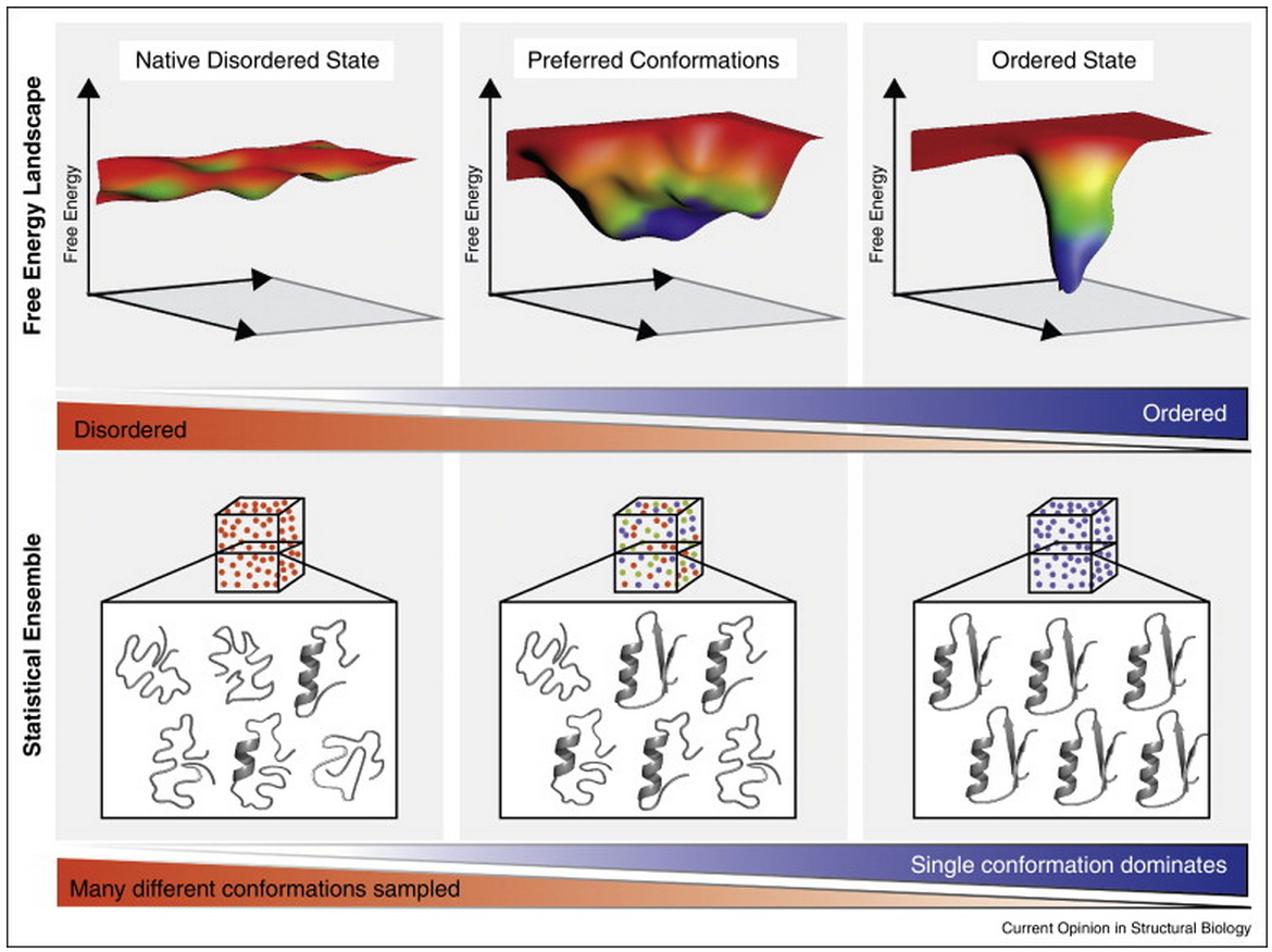
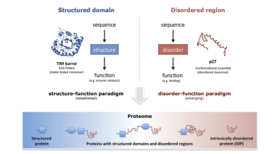
Intrinsically disordered regions (IDRs) are fundamental units of protein function and regulation. Despite their inability to form a unique stable tertiary structure in isolation, many IDRs adopt a defined conformation upon binding and achieve their function through their interactions with other biomolecules. However, this requirement for IDR functionality seems to be at odds with the high entropic cost they must incur upon binding an interaction partner. How is this seeming paradox resolved? While increasing the enthalpy of binding is one approach to compensate for this entropic cost, growing evidence suggests that inherent features of IDRs, for instance repeating linear motifs, minimise the entropic cost of binding. Moreover, this control of entropic cost can be carefully modulated by a range of regulatory mechanisms, such as alternative splicing and post-translational modifications, which enable allosteric communication and rheostat-like tuning of IDR function. In that sense, the high entropic cost of IDR binding can be advantageous by providing tunability to protein function. In addition to biological regulatory mechanisms, modulation of entropy can also be controlled by environmental factors, such as changes in temperature, redox-potential and pH. These principles are extensively exploited by a number of organisms, including pathogens. They can also be utilised in bioengineering, synthetic biology and in pharmaceutical applications such as increasing bioavailability of protein therapeutics. The review by Tilman Flock, Robert J Weatheritt, Natasha S Latysheva and M Madan Babu can be found here.

Structural polymorphism in the N-terminal oligomerization domain of NPM1

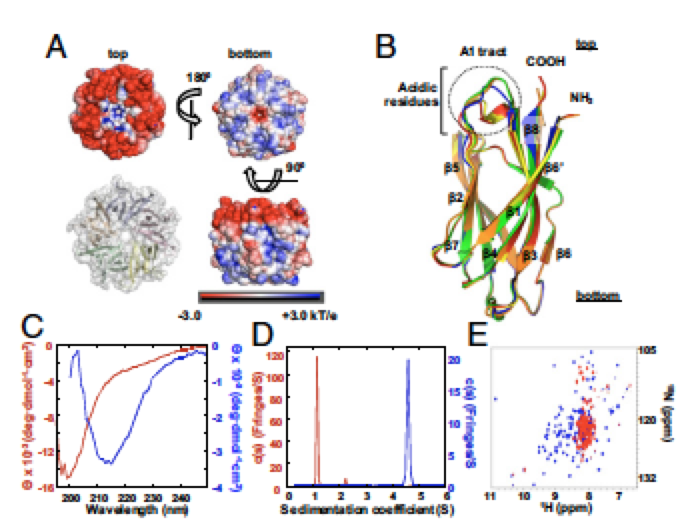
Nucleophosmin (NPM1) is a multifunctional phospho-protein with critical roles in ribosome biogenesis, tumor suppression, and nucleolar stress response. Here we show that the N-terminal oligomerization domain of NPM1 (Npm-N) exhibits structural polymorphism by populating conformational states ranging from a highly ordered, folded pentamer to a highly disordered monomer. The monomer–pentamer equilibrium is modulated by posttranslational modification and protein binding. Phosphorylation drives the equilibrium in favor of monomeric forms, and this effect can be reversed by Npm-N binding to its interaction partners. We have identified a short, arginine-rich linear motif in NPM1 binding partners that mediates Npm-N oligomerization. We propose that the diverse functional repertoire associated with NPM1 is controlled through a regulated unfolding mechanism signaled through posttranslational modifications and intermolecular interactions. The paper with Marija Buljan et al can be found here.

Our perspective on the hidden codes that constrain protein evolution is published in Science

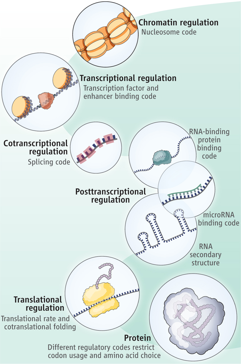
In this essay, we discuss how regulatory elements within protein-coding regions (such as transcription factor binding) can influence codon choice and amino acid preference that are independent of protein structure or function. We discuss how there may be conflicts between codes and highlight that the redundancy in the genetic code might facilitate the existence of multiple overlapping regulatory codes within protein-coding regions of the genome. You can read the Perspective here.


Promiscuity as a functional trait: intrinsically disordered regions as central players of interactomes

Because of their pervasiveness in eukaryotic genomes and their unique properties, understanding the role that ID (intrinsically disordered) regions in proteins play in the interactome is essential for gaining a better understanding of the network. Especially critical in determining this role is their ability to bind more than one partner using the same region. Studies have revealed that proteins containing ID regions tend to take a central role in protein interaction networks; specifically, they act as hubs, interacting with multiple different partners across time and space, allowing for the co-ordination of many cellular activities. There appear to be three different modules within ID regions responsible for their functionally promiscuous behaviour: MoRFs (molecular recognition features), SLiMs (small linear motifs) and LCRs (low complexity regions). These regions allow for functionality such as engaging in the formation of dynamic heteromeric structures which can serve to increase local activity of an enzyme or store a collection of functionally related molecules for later use. However, the use of promiscuity does not come without a cost: a number of diseases that have been associated with ID-containing proteins seem to be caused by undesirable interactions occurring upon altered expression of the ID-containing protein. The paper can be found here.


A methodology to infer gene networks from spatial patterns of expression – an application to fluorescence in situ hybridization images

The proper functional development of a multicellular organism depends on an intricate network of interacting genes that are expressed in accurate temporal and spatial patterns across different tissues. Complex inhibitory and excitatory interactions among genes control the territorial differences that explain specialized cell fates, embryo polarization and tissues architecture in metazoans. Given the nature of the regulatory gene networks, similarity of expression patterns can identify genes with similar roles. The inference and analysis of the gene interaction networks through complex network tools can reveal important aspects of the biological system modeled. Here we suggest an image analysis pipeline to quantify co-localization patterns in in situ hybridization images of Drosophila embryos and, based on these patterns, infer gene networks. We analyze the spatial dispersion of the gene expression and show the gene interaction networks for different developmental stages. Our results suggest that the inference of developmental networks based on spatial expression data is biologically relevant and represents a potential tool for the understanding of animal development. The paper can be found here.


Categories

Archives