Regulatory Genomics and Systems Biology

Proteome response at the edge of protein aggregation

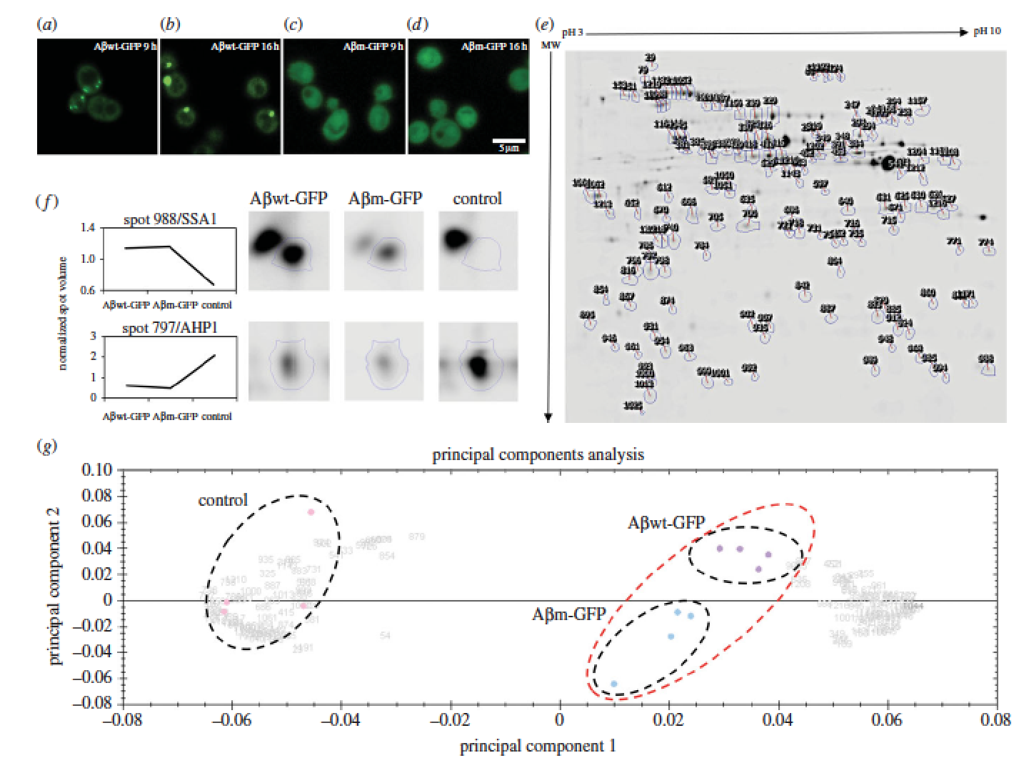
Proteins adopt defined structures and are crucial to most cellular functions. Their misfolding and aggregation is associated with numerous degenerative human disorders such as type II diabetes, Huntington’s or Alzheimer’s diseases. Here, we aim to understand why cells promote the formation of protein foci. Comparison of two amyloid-b-peptide variants, mostly insoluble but differently recruited by the cell (inclusion body versus diffused), reveals small differences in cell fitness and proteome response. We suggest that the levels of oxidative stress act as a sensor to trigger protein recruitment into foci. Our data support a common cytoplasmic response being able to discern and react to the specific properties of polypeptides. The paper by Natalia Sanchez de Groot can be viewed here.
Structured and disordered facets of the GPCR fold

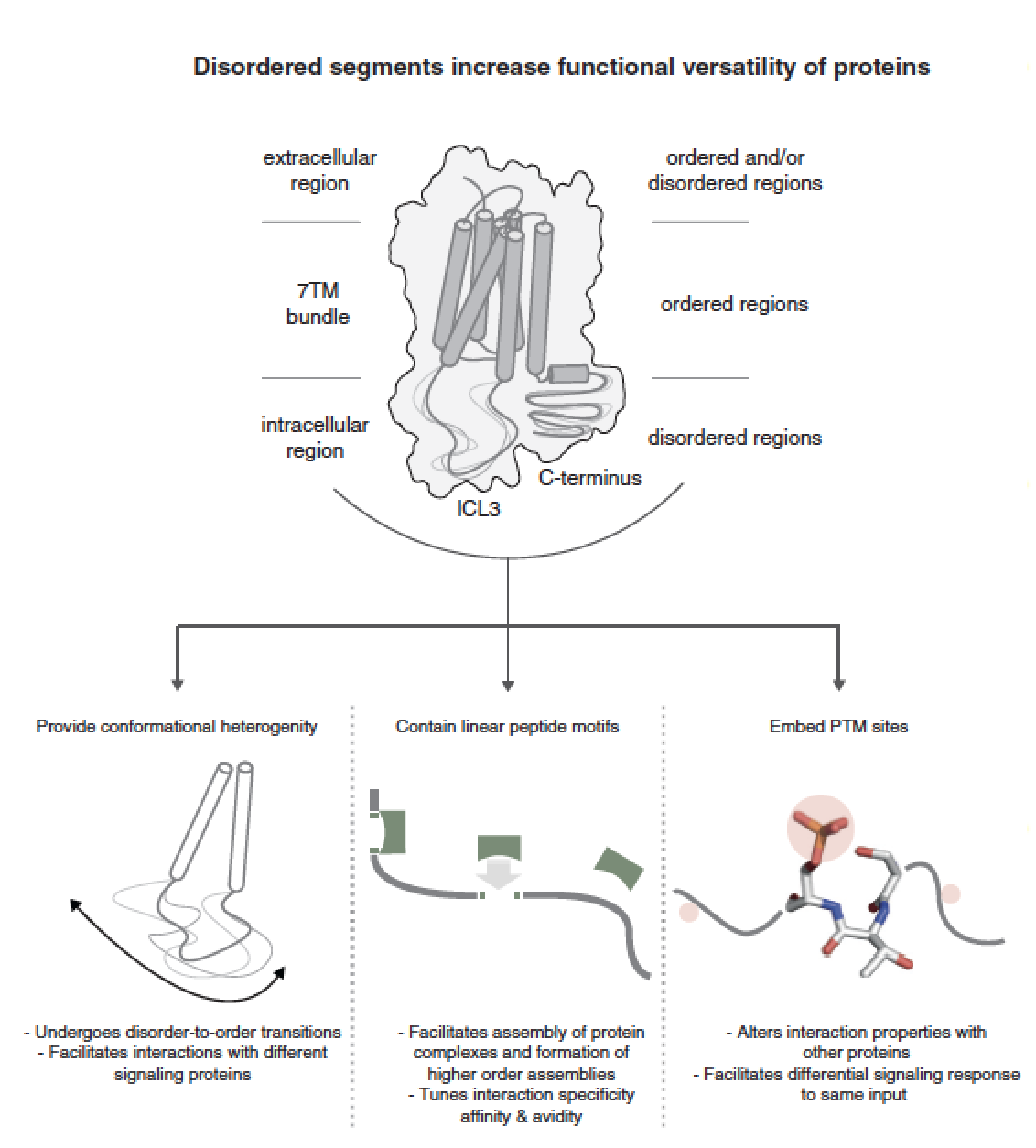
The seven-transmembrane (7TM) helix fold of G-protein coupled receptors (GPCRs) has been adapted for a wide variety of physiologically important signaling functions. Here, we discuss the diversity in the structured and disordered regions of GPCRs based on the recently published crystal structures and sequence analysis of all human GPCRs. A comparison of the structures of rhodopsin-like receptors (class A), secretin-like receptors (class B), metabotropic receptors (class C) and frizzled receptors (class F) shows that the relative arrangement of the transmembrane helices is conserved across all four GPCR classes although individual receptors can be activated by ligand binding at varying positions within and around the transmembrane helical bundle. A systematic analysis of GPCR sequences reveals the presence of disordered segments in the cytoplasmic side, abundant post-translational modification sites, evidence for alternative splicing and several putative linear peptide motifs that have the potential to mediate interactions with cytosolic proteins. While the structured regions permit the receptor to bind diverse ligands, the disordered regions appear to have an underappreciated role in modulating downstream signaling in response to the cellular state. An integrated paradigm combining the knowledge of structured and disordered regions is imperative for gaining a holistic understanding of the GPCR (un)structure-function relationship. The paper by AJ Venkatakrishnan et al can be viewed here.

Intrinsically Disordered Segments Affect Protein Half-Life in the Cell and during Evolution

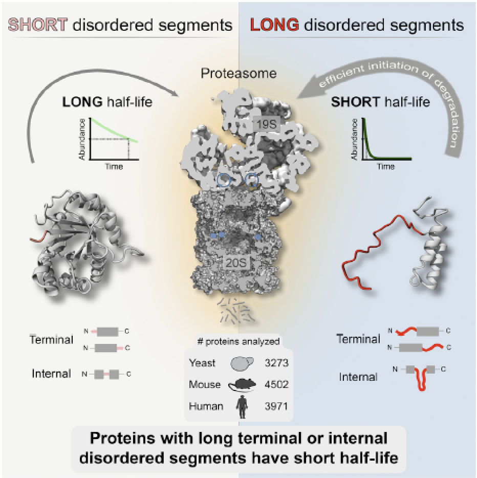
Precise control of protein turnover is essential for cellular homeostasis. The ubiquitin-proteasome system is well established as a major regulator of protein degradation, but an understanding of how inherent structural features influence the lifetimes of proteins is lacking. We report that yeast, mouse, and human proteins with terminal or internal intrinsically disordered segments have significantly shorter half-lives than proteins without these features. The lengths of the disordered segments that affect protein half-life are compatible with the structure of the proteasome. Divergence in terminal and internal disordered segments in yeast proteins originating from gene duplication leads to significantly altered half-life. Many paralogs that are affected by such changes participate in signaling, where altered protein half-life will directly impact cellular processes and function. Thus, natural variation in the length and position of disordered segments may affect protein half-life and could serve as an underappreciated source of genetic variation with important phenotypic consequences. The paper by Robin van der Lee et al can be found here.

Dr M. Madan Babu awarded Lister Research Prize

Congratulations to Madan to be awarded the prestigious Lister Research prize this year along with Melina Schuh, another LMB group leader. Madan’s Lister Research Prize has been awarded for his work on dynamics of tRNA abundance and the regulation of protein expression levels. He will deliver his Lister Prize Lecture at the awards ceremony on Friday 5th September 2014. Here is the detailed information as shown in the LMB website.

Asymmetric mRNA localization contributes to fidelity and sensitivity of spatially localized systems

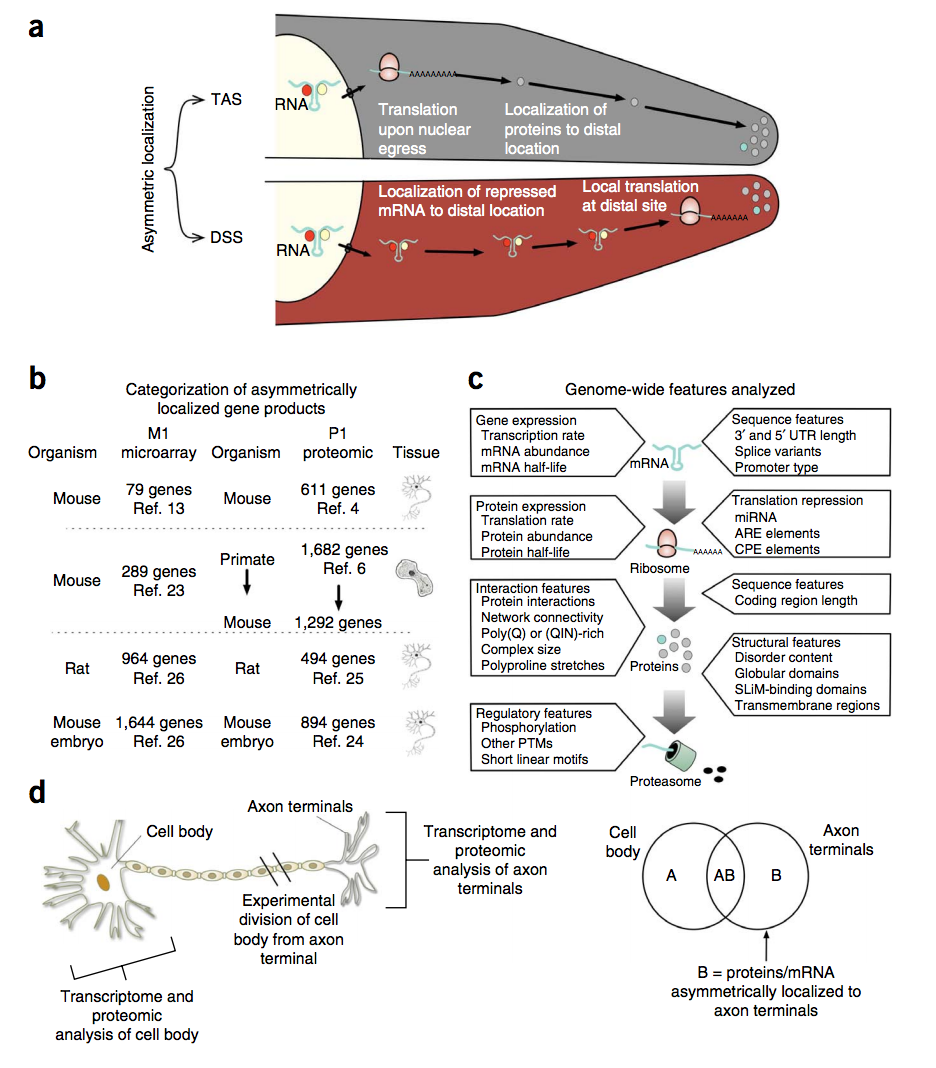
Although many proteins are localized after translation, asymmetric protein distribution is also achieved by translation after mRNA localization. Why are certain mRNA transported to a distal location and translated on-site? Here we undertake a systematic, genome-scale study of asymmetrically distributed protein and mRNA in mammalian cells. Our findings suggest that asymmetric protein distribution by mRNA localization enhances interaction fidelity and signaling sensitivity. Proteins synthesized at distal locations frequently contain intrinsically disordered segments. These regions are generally rich in assembly-promoting modules and are often regulated by post-translational modifications. Such proteins are tightly regulated but display distinct temporal dynamics upon stimulation with growth factors. Thus, proteins synthesized on-site may rapidly alter proteome composition and act as dynamically regulated scaffolds to promote the formation of reversible cellular assemblies. Our observations are consistent across multiple mammalian species, cell types and developmental stages, suggesting that localized translation is a recurring feature of cell signaling and regulation. The paper by Robert Weatheritt et al can be found here.

A Million Peptide Motifs for the Molecular Biologist

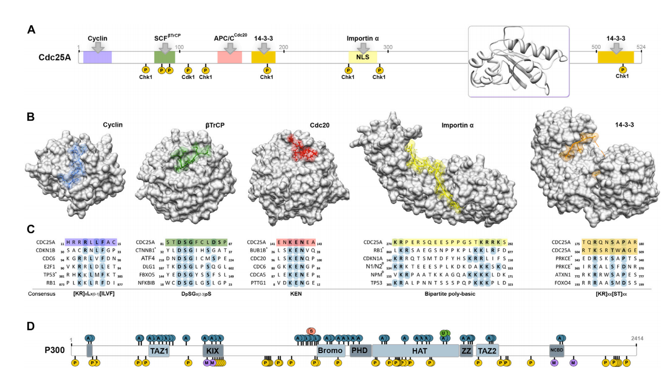
A molecular description of functional modules in the cell is the focus of many high-throughput studies in the postgenomic era. A large portion of biomolecular interactions in virtually all cellular processes is mediated by compact interaction modules, referred to as peptide motifs. Such motifs are typically less than ten residues in length, occur within intrinsically disordered regions, and are recognized and/or posttranslationally modified by structured domains of the interacting partner. In this review, we suggest that there might be over a million instances of peptide motifs in the human proteome. While this staggering number suggests that peptide motifs are numerous and the most understudied functional module in the cell, it also holds great opportunities for new discoveries. The review can be found here.